Home » Without Label » Blood Vessels Labeled - Quantification Of The Number Of Blood Vessels Labeled With Fluorescent Download Scientific Diagram - Learn vocabulary, terms, and more with flashcards, games, and other study tools.
Blood Vessels Labeled - Quantification Of The Number Of Blood Vessels Labeled With Fluorescent Download Scientific Diagram - Learn vocabulary, terms, and more with flashcards, games, and other study tools.
Blood Vessels Labeled - Quantification Of The Number Of Blood Vessels Labeled With Fluorescent Download Scientific Diagram - Learn vocabulary, terms, and more with flashcards, games, and other study tools.. Arteries are a type of blood vessel. The inferior vena cava is labeled in the figure below. Blood is supplied to parts within the neck, head and brain through branches of the subclavian and common carotid arteries. Blood vessels carry blood from your heart to the rest of your body. Like arteries, veins form a complex, branching system of larger and smaller vessels.
Use key choices to identify the blood vessel tunic described. Eventually, the smallest arteries, vessels called arterioles, further branch into tiny capillaries, where nutrients and wastes are exchanged. A primary purpose and significant role of the vasculature is its participation in oxygenating the body. The adventitia or outer layer which provides structural support and shape to the vessel This article lists a series of labeled imaging anatomy cases by system and modality.
Pin On A P 4 Heart Lung from i.pinimg.com Eventually, the smallest arteries, vessels called arterioles, further branch into tiny capillaries, where nutrients and wastes are exchanged. The smallest veins are called venules. Abstract—the segmentation of retinal blood vessels in the retina is a critical step in diagnosis of diabetic retinopathy. Start studying blood vessel anatomy. Learn vocabulary, terms, and more with flashcards, games, and other study tools. The common cartoid artery extends from the brachiocephalic artery. Veins (in blue) are the blood vessels that return blood to the heart. Capillaries come together to form venules, small blood vessels that carry blood to a vein, a larger blood vessel that returns blood to the heart.
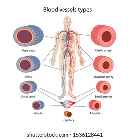
Blood vessels function to transport blood.in general, arteries and arterioles transport oxygenated blood from the lungs to the body and its organs, and veins and venules transport deoxygenated blood from the body to the lungs.blood vessels also circulate blood throughout the circulatory system oxygen (bound to hemoglobin in red blood cells) is the most critical nutrient carried by the blood.
Human heart labeling 27p image quiz. Arteries usually colored red because oxygen rich, carry blood away from the heart to capillaries within the tissues. Use key choices to identify the blood vessel tunic described. Arteries, veins, and capillaries blood vessels flow blood throughout the body. Blood vessels are divided into two broad categories: As the abdomen and pelvis contain the majority of internal organs, these regions need to be supplied by an extensive network of arteries and veins. They're essential for making sure your organs and tissues get the oxygen and nutrients they need to work. Abstract—the segmentation of retinal blood vessels in the retina is a critical step in diagnosis of diabetic retinopathy. 15 minutes it would be impossible to get blood to the predestined locations without the vascular pathways. Blood vessel labeling 7p image quiz. Disclosure • the material and the illustrations are adopted from the textbook human anatomy and physiology / ninth edition/ Dr calum worsley and assoc prof craig hacking et al. Capillaries are the smallest blood vessels where molecules move between blood and interstitial fluid of the tissues.
The common cartoid artery extends from the brachiocephalic artery. Abstract—the segmentation of retinal blood vessels in the retina is a critical step in diagnosis of diabetic retinopathy. Liver anatomy blood supply 19 photos of the liver anatomy blood supply anatomical location of liver, blood vessels that carry blood to the liver, dual blood supply to liver, functional anatomy of liver, liver and its functions, liver on the human body, normal anatomy of the liver, position of liver, inner body, anatomical location of … Capillaries come together to form venules, small blood vessels that carry blood to a vein, a larger blood vessel that returns blood to the heart. The microvasculature is composed of blood vessels that are smaller than 100 microns may only be seen through the microscope.
Chapter 19 Blood Vessels from image.slidesharecdn.com Vessels transport nutrients to organs/tissues and to transport wastes away from organs/tissues in the blood. Blood vessel labeling 9p image quiz. You can imagine the aorta and ivc as the two trees, with all. Dimitrios mytilinaios md, phd last reviewed: Once blood is oxygenated in the lungs, it returns to the heart and is then pumped throughout the body. These vessels connect other organs in your body to your heart. Dr calum worsley and assoc prof craig hacking et al. Blood vessels form the extensive networks by which blood leaves the heart to supply tissue.
In contrast, veins carry blood back to the heart.
Blood is circulated through the body by blood vessels via the cardiovascular system which is comprised of the heart and the circulatory system.arteries move blood from the heart first to smaller arterioles, then capillaries or sinusoids, venules, veins, and back to the heart. Anatomy of blood vessels review sheet 32 261 microscopic structure of the blood vessels 1. You can imagine the aorta and ivc as the two trees, with all. Because arteries are moving blood being pumped out by the heart. Deoxygenated blood from the peripheral veins is transported back to the heart from capillaries, to venules, to veins, to the right side of the heart, and then. Eventually, the smallest arteries, vessels called arterioles, further branch into tiny capillaries, where nutrients and wastes are exchanged. Arteries (in red) are the blood vessels that deliver blood to the body. They work to carry blood away from the heart. The vessels that carry blood away from the heart are called arteries, and their very small branches are arterioles. A web of blood vessels—arteries, veins, and capillaries—circulate blood to organs. Once blood is oxygenated in the lungs, it returns to the heart and is then pumped throughout the body. Very small branches that collect the blood from the various organs and parts are called venules, and they unite to form veins, which return the blood to the heart. In contrast, veins carry blood back to the heart.
These vessels connect other organs in your body to your heart. The function and structure of each segment of the peripheral vascular system vary depending on the organ it supplies. Blood is circulated through the body by blood vessels via the cardiovascular system which is comprised of the heart and the circulatory system.arteries move blood from the heart first to smaller arterioles, then capillaries or sinusoids, venules, veins, and back to the heart. But blood vessels can develop problems, such as blockages or enlargement. Dimitrios mytilinaios md, phd last reviewed:
Arteries Veins Capillaries Diagram High Res Stock Images Shutterstock from image.shutterstock.com External veins and arteries of the heart ec by mrsdohm 65,191 plays 8p image quiz. Eventually, the smallest arteries, vessels called arterioles, further branch into tiny capillaries, where nutrients and wastes are exchanged, and then combine with other vessels that exit capillaries to form venules, small blood vessels that carry blood to a vein, a larger blood vessel that returns blood to the heart. A web of blood vessels—arteries, veins, and capillaries—circulate blood to organs. 15 minutes it would be impossible to get blood to the predestined locations without the vascular pathways. They work to carry blood away from the heart. The function and structure of each segment of the peripheral vascular system vary depending on the organ it supplies. Use key choices to identify the blood vessel tunic described. Learn vocabulary, terms, and more with flashcards, games, and other study tools.
Blood vessel labeling 7p image quiz.
Lorenzo crumbie mbbs, bsc • reviewer: Blood vessels are divided into two broad categories: But blood vessels can develop problems, such as blockages or enlargement. Human heart labeling 27p image quiz. Very small branches that collect the blood from the various organs and parts are called venules, and they unite to form veins, which return the blood to the heart. Blood cells by descartes 48,965 plays 9p image quiz. The three major types of blood vessels: Eventually, the smallest arteries, vessels called arterioles, further branch into tiny capillaries, where nutrients and wastes are exchanged, and then combine with other vessels that exit capillaries to form venules, small blood vessels that carry blood to a vein, a larger blood vessel that returns blood to the heart. Start studying blood vessels labeling. Arteries (in red) are the blood vessels that deliver blood to the body. Blood vessels carry blood from your heart to the rest of your body. Abstract—the segmentation of retinal blood vessels in the retina is a critical step in diagnosis of diabetic retinopathy. The venules and veins returning blood to the heart.